Usual questions
from our patients

- What is an Echocardiogram?
- Why do I need an Echocardiogram?
- Does an Echocardiogram use radiation?
- Is it a painful procedure?
- What Does Echocardiography Show?
- Is there special preparation needed?
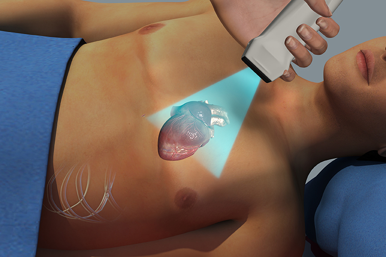
Echocardiogram is an ultrasound examination of the heart. An echocardiogram is a noninvasive (the skin is not pierced) procedure used to assess the heart’s function and structures. During the procedure, a transducer (like a microphone) sends out sound waves at a frequency too high to be heard. Sound waves are used to make a picture of your heart.
An echocardiogram allows the doctor to see different parts of your heart and how well they are working.
An echocardiogram is a test that uses sound waves to create pictures of the heart. The picture and information it produces is more detailed than a standard x-ray image. An echocardiogram does not expose you to radiation
This imaging procedure is not invasive and carries little to no risks. You may have discomfort from the positioning of the transducer because it can put pressure on the surface of the body.
Echocardiography (echo) shows the size, structure, and movement of various parts of your heart. These parts include the heart valves, the septum (the wall separating the right and left heart chambers), and the walls of the heart chambers. Doppler ultrasound shows the movement of blood through your heart.
Generally, you don’t need to do any preparation such as fasting or having sedation.
Tell your doctor of all prescription and over-the-counter medicines and herbal supplements that you are taking.
Tell your doctor if you have a pacemaker.
- Will i have anesthesia?
- What should I wear for the test?
- How long does the test take to complete?
- What To Expect During Echocardiography?
- What To Expect After Echocardiography?
- When will you know the results of your test?
No anesthesia will be used
Wear a shirt or blouse that can be taken off easily. Females will be asked to remove their bras. All patients will be given a gown to wear during the test.
The test will take about 40 to 50 minutes to complete.
During the test you will be lying down on your left side.
First You will be connected to an ECG machine that records the electrical activity of the heart and monitors the heart during the procedure using small, adhesive electrodes. The ECG tracings that record the electrical activity of the heart will be compared with the images displayed on the echocardiogram monitor.
The room will be darkened so that the images on the echo monitor can be seen by the sonographer (ultrasound tech).
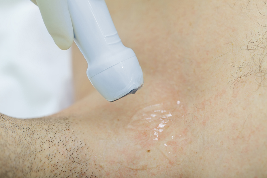
A special gel will be placed on the front of your chest and then the sonographer will take a special probe and move it around your chest area, applying varying amounts of pressure to get images of different locations and structures of your heart. This will make a picture of your heart that can be seen on a monitor.
After the procedure, the technologist will wipe the gel from your chest and remove the ECG electrode pads. You may then put on your clothes.
You usually can go back to your normal activities right after having echocardiography (echo).
You may resume your usual diet and life style unless your doctor tells you differently.
The results of the test will be sent to the doctor that ordered the test for you.
It may take 3 to 4 days before you know the results of your test.
Please note: The sonographer that is doing your echocardiogram will not give you the results of the test after the procedure
Please contact your cardiologists for the results.


